Vanadium »
PDB 4zi4-6py9 »
6db2 »
Vanadium in PDB 6db2: X-Ray Crystal Structure of Vioc Bound to Vanadyl Ion, L-Homoarginine, and Succinate
Enzymatic activity of X-Ray Crystal Structure of Vioc Bound to Vanadyl Ion, L-Homoarginine, and Succinate
All present enzymatic activity of X-Ray Crystal Structure of Vioc Bound to Vanadyl Ion, L-Homoarginine, and Succinate:
1.14.11.41;
1.14.11.41;
Protein crystallography data
The structure of X-Ray Crystal Structure of Vioc Bound to Vanadyl Ion, L-Homoarginine, and Succinate, PDB code: 6db2
was solved by
N.P.Dunham,
A.J.Mitchell,
A.K.Boal,
with X-Ray Crystallography technique. A brief refinement statistics is given in the table below:
| Resolution Low / High (Å) | 59.61 / 1.70 |
| Space group | C 1 2 1 |
| Cell size a, b, c (Å), α, β, γ (°) | 81.025, 66.637, 63.224, 90.00, 109.47, 90.00 |
| R / Rfree (%) | 17.5 / 20 |
Vanadium Binding Sites:
The binding sites of Vanadium atom in the X-Ray Crystal Structure of Vioc Bound to Vanadyl Ion, L-Homoarginine, and Succinate
(pdb code 6db2). This binding sites where shown within
5.0 Angstroms radius around Vanadium atom.
In total only one binding site of Vanadium was determined in the X-Ray Crystal Structure of Vioc Bound to Vanadyl Ion, L-Homoarginine, and Succinate, PDB code: 6db2:
In total only one binding site of Vanadium was determined in the X-Ray Crystal Structure of Vioc Bound to Vanadyl Ion, L-Homoarginine, and Succinate, PDB code: 6db2:
Vanadium binding site 1 out of 1 in 6db2
Go back to
Vanadium binding site 1 out
of 1 in the X-Ray Crystal Structure of Vioc Bound to Vanadyl Ion, L-Homoarginine, and Succinate
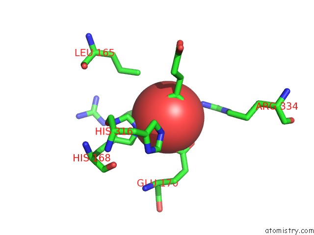
Mono view
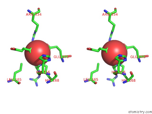
Stereo pair view
Mono view
Stereo pair view
A full contact list of Vanadium with other atoms in the V binding
site number 1 of X-Ray Crystal Structure of Vioc Bound to Vanadyl Ion, L-Homoarginine, and Succinate within 5.0Å range:
|
Reference:
N.P.Dunham,
W.C.Chang,
A.J.Mitchell,
R.J.Martinie,
B.Zhang,
J.A.Bergman,
L.J.Rajakovich,
B.Wang,
A.Silakov,
C.Krebs,
A.K.Boal,
J.M.Bollinger.
Two Distinct Mechanisms For C-C Desaturation By Iron(II)- and 2-(Oxo)Glutarate-Dependent Oxygenases: Importance of Alpha-Heteroatom Assistance. J. Am. Chem. Soc. V. 140 7116 2018.
ISSN: ESSN 1520-5126
PubMed: 29708749
DOI: 10.1021/JACS.8B01933
Page generated: Fri Oct 11 20:02:30 2024
ISSN: ESSN 1520-5126
PubMed: 29708749
DOI: 10.1021/JACS.8B01933
Last articles
Mg in 3WYFMg in 3WYG
Mg in 3WY3
Mg in 3WY4
Mg in 3WY2
Mg in 3WY1
Mg in 3WXM
Mg in 3WXX
Mg in 3WXL
Mg in 3WVE