Vanadium »
PDB 1b8j-1yv3 »
1ruv »
Vanadium in PDB 1ruv: Ribonuclease A-Uridine Vanadate Complex: High Resolution Resolution X-Ray Structure (1.3 A)
Enzymatic activity of Ribonuclease A-Uridine Vanadate Complex: High Resolution Resolution X-Ray Structure (1.3 A)
All present enzymatic activity of Ribonuclease A-Uridine Vanadate Complex: High Resolution Resolution X-Ray Structure (1.3 A):
3.1.27.5;
3.1.27.5;
Protein crystallography data
The structure of Ribonuclease A-Uridine Vanadate Complex: High Resolution Resolution X-Ray Structure (1.3 A), PDB code: 1ruv
was solved by
J.E.Ladner,
B.Wladkowski,
L.A.Svensson,
L.Sjolin,
G.L.Gilliland,
with X-Ray Crystallography technique. A brief refinement statistics is given in the table below:
| Resolution Low / High (Å) | 10.00 / 1.25 |
| Space group | P 1 21 1 |
| Cell size a, b, c (Å), α, β, γ (°) | 29.800, 38.200, 53.200, 90.00, 106.10, 90.00 |
| R / Rfree (%) | n/a / n/a |
Vanadium Binding Sites:
The binding sites of Vanadium atom in the Ribonuclease A-Uridine Vanadate Complex: High Resolution Resolution X-Ray Structure (1.3 A)
(pdb code 1ruv). This binding sites where shown within
5.0 Angstroms radius around Vanadium atom.
In total only one binding site of Vanadium was determined in the Ribonuclease A-Uridine Vanadate Complex: High Resolution Resolution X-Ray Structure (1.3 A), PDB code: 1ruv:
In total only one binding site of Vanadium was determined in the Ribonuclease A-Uridine Vanadate Complex: High Resolution Resolution X-Ray Structure (1.3 A), PDB code: 1ruv:
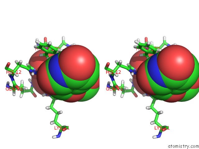
Vanadium binding site 1 out of 1 in 1ruv
Go back to
Vanadium binding site 1 out
of 1 in the Ribonuclease A-Uridine Vanadate Complex: High Resolution Resolution X-Ray Structure (1.3 A)
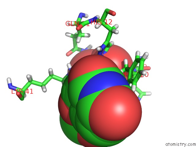
Mono view
Stereo pair view
Mono view
Stereo pair view
A full contact list of Vanadium with other atoms in the V binding
site number 1 of Ribonuclease A-Uridine Vanadate Complex: High Resolution Resolution X-Ray Structure (1.3 A) within 5.0Å range:
|
Reference:
J.E.Ladner,
B.D.Wladkowski,
L.A.Svensson,
L.Sjolin,
G.L.Gilliland.
X-Ray Structure of A Ribonuclease A-Uridine Vanadate Complex at 1.3 A Resolution. Acta Crystallogr.,Sect.D V. 53 290 1997.
ISSN: ISSN 0907-4449
PubMed: 15299932
DOI: 10.1107/S090744499601582X
Page generated: Tue Aug 19 07:08:56 2025
ISSN: ISSN 0907-4449
PubMed: 15299932
DOI: 10.1107/S090744499601582X
Last articles
K in 9CWUK in 9CVB
K in 9CVA
K in 9COM
Fe in 9VR0
Fe in 9UD8
Fe in 9QDT
Fe in 9S2T
Fe in 9JQA
Fe in 9IYV