Vanadium »
PDB 7q0v-9g2m »
7q0w »
Vanadium in PDB 7q0w: Bovine Trypsin Co-Crystallized with V(IV)OSO4 and Phen
Enzymatic activity of Bovine Trypsin Co-Crystallized with V(IV)OSO4 and Phen
All present enzymatic activity of Bovine Trypsin Co-Crystallized with V(IV)OSO4 and Phen:
3.4.21.4;
3.4.21.4;
Protein crystallography data
The structure of Bovine Trypsin Co-Crystallized with V(IV)OSO4 and Phen, PDB code: 7q0w
was solved by
M.F.A.Santos,
A.C.P.Fernandes,
I.Correia,
G.Sciortino,
E.Garribba,
T.Santos-Silva,
J.C.Pessoa,
with X-Ray Crystallography technique. A brief refinement statistics is given in the table below:
| Resolution Low / High (Å) | 42.68 / 1.20 |
| Space group | P 21 21 21 |
| Cell size a, b, c (Å), α, β, γ (°) | 53.905, 56.082, 65.779, 90, 90, 90 |
| R / Rfree (%) | 12.7 / 16.2 |
Other elements in 7q0w:
The structure of Bovine Trypsin Co-Crystallized with V(IV)OSO4 and Phen also contains other interesting chemical elements:
| Calcium | (Ca) | 1 atom |
Vanadium Binding Sites:
The binding sites of Vanadium atom in the Bovine Trypsin Co-Crystallized with V(IV)OSO4 and Phen
(pdb code 7q0w). This binding sites where shown within
5.0 Angstroms radius around Vanadium atom.
In total only one binding site of Vanadium was determined in the Bovine Trypsin Co-Crystallized with V(IV)OSO4 and Phen, PDB code: 7q0w:
In total only one binding site of Vanadium was determined in the Bovine Trypsin Co-Crystallized with V(IV)OSO4 and Phen, PDB code: 7q0w:
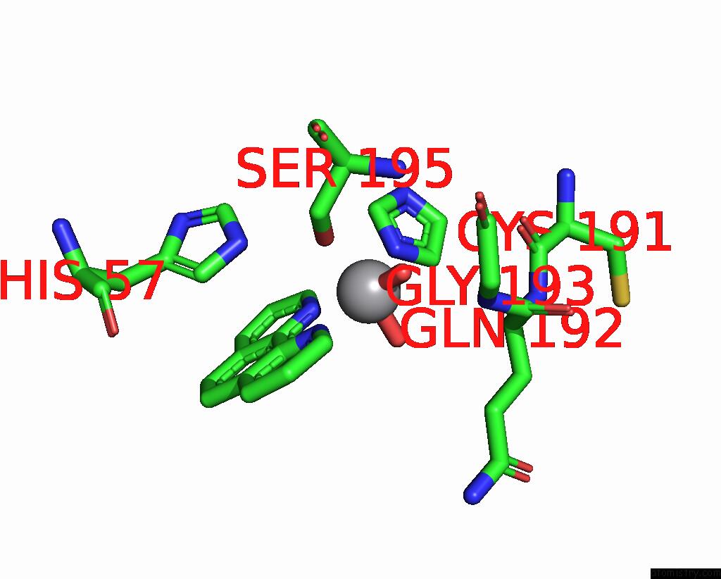
Vanadium binding site 1 out of 1 in 7q0w
Go back to
Vanadium binding site 1 out
of 1 in the Bovine Trypsin Co-Crystallized with V(IV)OSO4 and Phen
Mono view

Stereo pair view
Mono view
Stereo pair view
A full contact list of Vanadium with other atoms in the V binding
site number 1 of Bovine Trypsin Co-Crystallized with V(IV)OSO4 and Phen within 5.0Å range:
|
Reference:
M.F.A.Santos,
G.Sciortino,
I.Correia,
A.C.P.Fernandes,
T.Santos-Silva,
F.Pisanu,
E.Garribba,
J.Costa Pessoa.
Binding of V IV O 2+ , V IV Ol, V IV Ol 2 and V V O 2 L Moieties to Proteins: X-Ray/Theoretical Characterization and Biological Implications. Chemistry V. 28 00105 2022.
ISSN: ISSN 0947-6539
PubMed: 35486702
DOI: 10.1002/CHEM.202200105
Page generated: Fri Oct 11 20:41:16 2024
ISSN: ISSN 0947-6539
PubMed: 35486702
DOI: 10.1002/CHEM.202200105
Last articles
Zn in 9MJ5Zn in 9HNW
Zn in 9G0L
Zn in 9FNE
Zn in 9DZN
Zn in 9E0I
Zn in 9D32
Zn in 9DAK
Zn in 8ZXC
Zn in 8ZUF