Vanadium »
PDB 4zi4-6py9 »
6phs »
Vanadium in PDB 6phs: Protein Tyrosine Phosphatase 1B (1-301), P185A Mutant, Vanadate Bound State
Enzymatic activity of Protein Tyrosine Phosphatase 1B (1-301), P185A Mutant, Vanadate Bound State
All present enzymatic activity of Protein Tyrosine Phosphatase 1B (1-301), P185A Mutant, Vanadate Bound State:
3.1.3.48;
3.1.3.48;
Protein crystallography data
The structure of Protein Tyrosine Phosphatase 1B (1-301), P185A Mutant, Vanadate Bound State, PDB code: 6phs
was solved by
D.S.Cui,
J.M.Lipchock,
J.P.Loria,
with X-Ray Crystallography technique. A brief refinement statistics is given in the table below:
| Resolution Low / High (Å) | 40.35 / 2.13 |
| Space group | P 31 2 1 |
| Cell size a, b, c (Å), α, β, γ (°) | 87.704, 87.704, 102.961, 90.00, 90.00, 120.00 |
| R / Rfree (%) | 19.8 / 24.1 |
Vanadium Binding Sites:
The binding sites of Vanadium atom in the Protein Tyrosine Phosphatase 1B (1-301), P185A Mutant, Vanadate Bound State
(pdb code 6phs). This binding sites where shown within
5.0 Angstroms radius around Vanadium atom.
In total only one binding site of Vanadium was determined in the Protein Tyrosine Phosphatase 1B (1-301), P185A Mutant, Vanadate Bound State, PDB code: 6phs:
In total only one binding site of Vanadium was determined in the Protein Tyrosine Phosphatase 1B (1-301), P185A Mutant, Vanadate Bound State, PDB code: 6phs:
Vanadium binding site 1 out of 1 in 6phs
Go back to
Vanadium binding site 1 out
of 1 in the Protein Tyrosine Phosphatase 1B (1-301), P185A Mutant, Vanadate Bound State
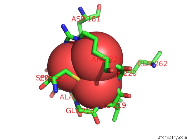
Mono view
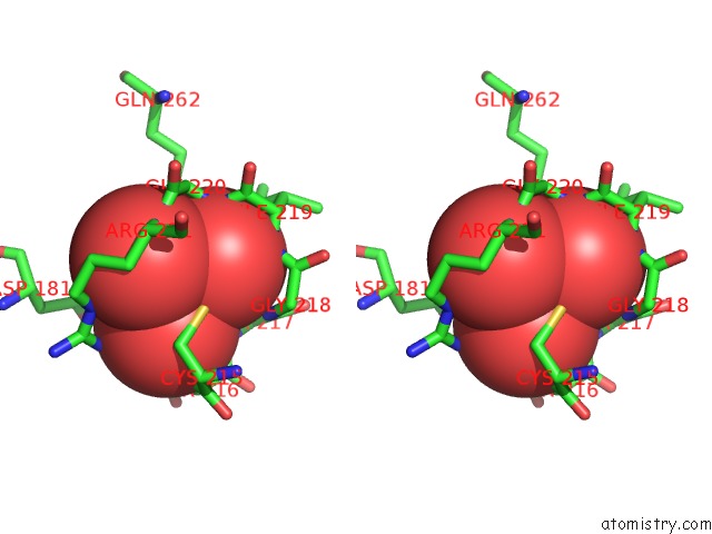
Stereo pair view
Mono view
Stereo pair view
A full contact list of Vanadium with other atoms in the V binding
site number 1 of Protein Tyrosine Phosphatase 1B (1-301), P185A Mutant, Vanadate Bound State within 5.0Å range:
|
Reference:
D.S.Cui,
J.M.Lipchock,
D.Brookner,
J.P.Loria.
Uncovering the Molecular Interactions in the Catalytic Loop That Modulate the Conformational Dynamics in Protein Tyrosine Phosphatase 1B. J.Am.Chem.Soc. V. 141 12634 2019.
ISSN: ESSN 1520-5126
PubMed: 31339043
DOI: 10.1021/JACS.9B04470
Page generated: Fri Oct 11 20:09:51 2024
ISSN: ESSN 1520-5126
PubMed: 31339043
DOI: 10.1021/JACS.9B04470
Last articles
Fe in 2YXOFe in 2YRS
Fe in 2YXC
Fe in 2YNM
Fe in 2YVJ
Fe in 2YP1
Fe in 2YU2
Fe in 2YU1
Fe in 2YQB
Fe in 2YOO