Vanadium »
PDB 4zi4-6py9 »
5ijs »
Vanadium in PDB 5ijs: Crystal Structure of Autotaxin with Orthovanadate Bound As A Trigonal Bipyramidal Intermediate Analog
Enzymatic activity of Crystal Structure of Autotaxin with Orthovanadate Bound As A Trigonal Bipyramidal Intermediate Analog
All present enzymatic activity of Crystal Structure of Autotaxin with Orthovanadate Bound As A Trigonal Bipyramidal Intermediate Analog:
3.1.4.39;
3.1.4.39;
Protein crystallography data
The structure of Crystal Structure of Autotaxin with Orthovanadate Bound As A Trigonal Bipyramidal Intermediate Analog, PDB code: 5ijs
was solved by
J.Hausmann,
R.P.Joosten,
A.Perrakis,
with X-Ray Crystallography technique. A brief refinement statistics is given in the table below:
| Resolution Low / High (Å) | 40.00 / 2.20 |
| Space group | P 1 |
| Cell size a, b, c (Å), α, β, γ (°) | 53.750, 63.450, 70.550, 99.33, 105.91, 99.51 |
| R / Rfree (%) | 20.3 / 23.9 |
Other elements in 5ijs:
The structure of Crystal Structure of Autotaxin with Orthovanadate Bound As A Trigonal Bipyramidal Intermediate Analog also contains other interesting chemical elements:
| Zinc | (Zn) | 2 atoms |
| Iodine | (I) | 6 atoms |
| Calcium | (Ca) | 1 atom |
| Sodium | (Na) | 2 atoms |
Vanadium Binding Sites:
The binding sites of Vanadium atom in the Crystal Structure of Autotaxin with Orthovanadate Bound As A Trigonal Bipyramidal Intermediate Analog
(pdb code 5ijs). This binding sites where shown within
5.0 Angstroms radius around Vanadium atom.
In total only one binding site of Vanadium was determined in the Crystal Structure of Autotaxin with Orthovanadate Bound As A Trigonal Bipyramidal Intermediate Analog, PDB code: 5ijs:
In total only one binding site of Vanadium was determined in the Crystal Structure of Autotaxin with Orthovanadate Bound As A Trigonal Bipyramidal Intermediate Analog, PDB code: 5ijs:
Vanadium binding site 1 out of 1 in 5ijs
Go back to
Vanadium binding site 1 out
of 1 in the Crystal Structure of Autotaxin with Orthovanadate Bound As A Trigonal Bipyramidal Intermediate Analog
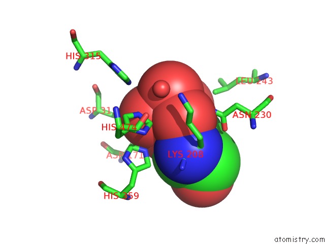
Mono view
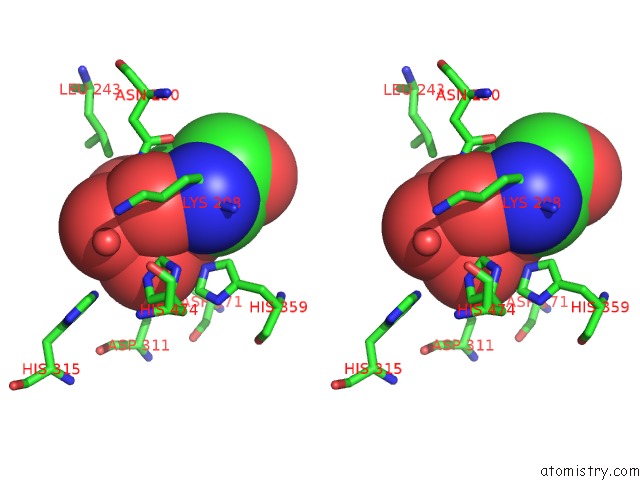
Stereo pair view
Mono view
Stereo pair view
A full contact list of Vanadium with other atoms in the V binding
site number 1 of Crystal Structure of Autotaxin with Orthovanadate Bound As A Trigonal Bipyramidal Intermediate Analog within 5.0Å range:
|
Reference:
J.Hausmann,
W.J.Keune,
A.L.Hipgrave Ederveen,
L.Van Zeijl,
R.P.Joosten,
A.Perrakis.
Structural Snapshots of the Catalytic Cycle of the Phosphodiesterase Autotaxin. J.Struct.Biol. V. 195 199 2016.
ISSN: ESSN 1095-8657
PubMed: 27268273
DOI: 10.1016/J.JSB.2016.06.002
Page generated: Fri Oct 11 19:59:16 2024
ISSN: ESSN 1095-8657
PubMed: 27268273
DOI: 10.1016/J.JSB.2016.06.002
Last articles
Cl in 5LE1Cl in 5LDR
Cl in 5LDP
Cl in 5LDQ
Cl in 5LAY
Cl in 5LDI
Cl in 5LDM
Cl in 5LDB
Cl in 5LD9
Cl in 5LCJ