Vanadium »
PDB 3omx-4zg4 »
4qih »
Vanadium in PDB 4qih: The Structure of Mycobacterial Glucosyl-3-Phosphoglycerate Phosphatase RV2419C Complexes with VO3
Enzymatic activity of The Structure of Mycobacterial Glucosyl-3-Phosphoglycerate Phosphatase RV2419C Complexes with VO3
All present enzymatic activity of The Structure of Mycobacterial Glucosyl-3-Phosphoglycerate Phosphatase RV2419C Complexes with VO3:
3.1.3.70;
3.1.3.70;
Protein crystallography data
The structure of The Structure of Mycobacterial Glucosyl-3-Phosphoglycerate Phosphatase RV2419C Complexes with VO3, PDB code: 4qih
was solved by
W.H.Zhou,
Q.Q.Zheng,
D.Q.Jiang,
W.Zhang,
Q.Q.Zhang,
J.Jin,
X.Li,
H.T.Yang,
N.Shaw,
Z.Rao,
with X-Ray Crystallography technique. A brief refinement statistics is given in the table below:
| Resolution Low / High (Å) | 35.01 / 2.30 |
| Space group | P 21 21 21 |
| Cell size a, b, c (Å), α, β, γ (°) | 46.312, 82.762, 131.319, 90.00, 90.00, 90.00 |
| R / Rfree (%) | 18.7 / 24.5 |
Vanadium Binding Sites:
The binding sites of Vanadium atom in the The Structure of Mycobacterial Glucosyl-3-Phosphoglycerate Phosphatase RV2419C Complexes with VO3
(pdb code 4qih). This binding sites where shown within
5.0 Angstroms radius around Vanadium atom.
In total 2 binding sites of Vanadium where determined in the The Structure of Mycobacterial Glucosyl-3-Phosphoglycerate Phosphatase RV2419C Complexes with VO3, PDB code: 4qih:
Jump to Vanadium binding site number: 1; 2;
In total 2 binding sites of Vanadium where determined in the The Structure of Mycobacterial Glucosyl-3-Phosphoglycerate Phosphatase RV2419C Complexes with VO3, PDB code: 4qih:
Jump to Vanadium binding site number: 1; 2;
Vanadium binding site 1 out of 2 in 4qih
Go back to
Vanadium binding site 1 out
of 2 in the The Structure of Mycobacterial Glucosyl-3-Phosphoglycerate Phosphatase RV2419C Complexes with VO3
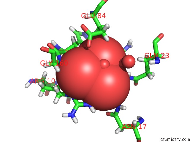
Mono view
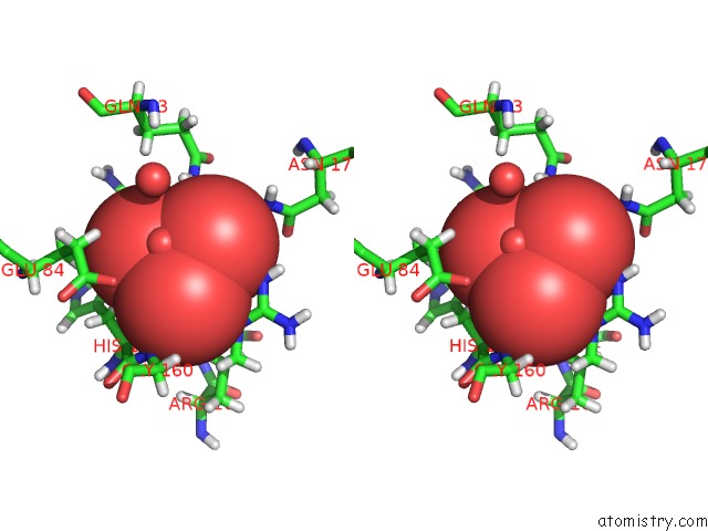
Stereo pair view
Mono view
Stereo pair view
A full contact list of Vanadium with other atoms in the V binding
site number 1 of The Structure of Mycobacterial Glucosyl-3-Phosphoglycerate Phosphatase RV2419C Complexes with VO3 within 5.0Å range:
|
Vanadium binding site 2 out of 2 in 4qih
Go back to
Vanadium binding site 2 out
of 2 in the The Structure of Mycobacterial Glucosyl-3-Phosphoglycerate Phosphatase RV2419C Complexes with VO3
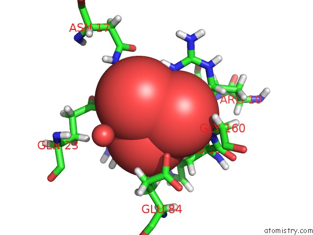
Mono view
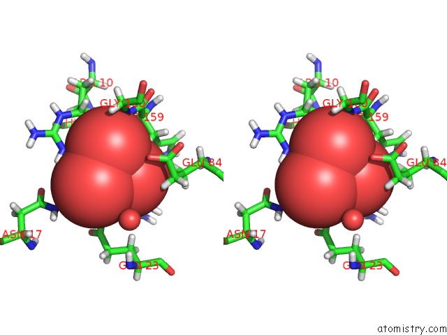
Stereo pair view
Mono view
Stereo pair view
A full contact list of Vanadium with other atoms in the V binding
site number 2 of The Structure of Mycobacterial Glucosyl-3-Phosphoglycerate Phosphatase RV2419C Complexes with VO3 within 5.0Å range:
|
Reference:
Q.Zheng,
D.Jiang,
W.Zhang,
Q.Zhang,
Q.Zhao,
J.Jin,
X.Li,
H.Yang,
M.Bartlam,
N.Shaw,
W.Zhou,
Z.Rao.
Mechanism of Dephosphorylation of Glucosyl-3-Phosphoglycerate By A Histidine Phosphatase J.Biol.Chem. V. 289 21242 2014.
ISSN: ISSN 0021-9258
PubMed: 24914210
DOI: 10.1074/JBC.M114.569913
Page generated: Fri Oct 11 19:44:35 2024
ISSN: ISSN 0021-9258
PubMed: 24914210
DOI: 10.1074/JBC.M114.569913
Last articles
Fe in 2YXOFe in 2YRS
Fe in 2YXC
Fe in 2YNM
Fe in 2YVJ
Fe in 2YP1
Fe in 2YU2
Fe in 2YU1
Fe in 2YQB
Fe in 2YOO