Vanadium »
PDB 3omx-4zg4 »
4erc »
Vanadium in PDB 4erc: Structure of Vhz Bound to Metavanadate
Enzymatic activity of Structure of Vhz Bound to Metavanadate
Protein crystallography data
The structure of Structure of Vhz Bound to Metavanadate, PDB code: 4erc
was solved by
K.Vyacheslav,
C.H.Alvan,
J.J.Sean,
with X-Ray Crystallography technique. A brief refinement statistics is given in the table below:
| Resolution Low / High (Å) | 31.32 / 1.15 |
| Space group | P 21 21 21 |
| Cell size a, b, c (Å), α, β, γ (°) | 32.992, 79.970, 99.718, 90.00, 90.00, 90.00 |
| R / Rfree (%) | 12.8 / 14.5 |
Vanadium Binding Sites:
The binding sites of Vanadium atom in the Structure of Vhz Bound to Metavanadate
(pdb code 4erc). This binding sites where shown within
5.0 Angstroms radius around Vanadium atom.
In total 2 binding sites of Vanadium where determined in the Structure of Vhz Bound to Metavanadate, PDB code: 4erc:
Jump to Vanadium binding site number: 1; 2;
In total 2 binding sites of Vanadium where determined in the Structure of Vhz Bound to Metavanadate, PDB code: 4erc:
Jump to Vanadium binding site number: 1; 2;
Vanadium binding site 1 out of 2 in 4erc
Go back to
Vanadium binding site 1 out
of 2 in the Structure of Vhz Bound to Metavanadate
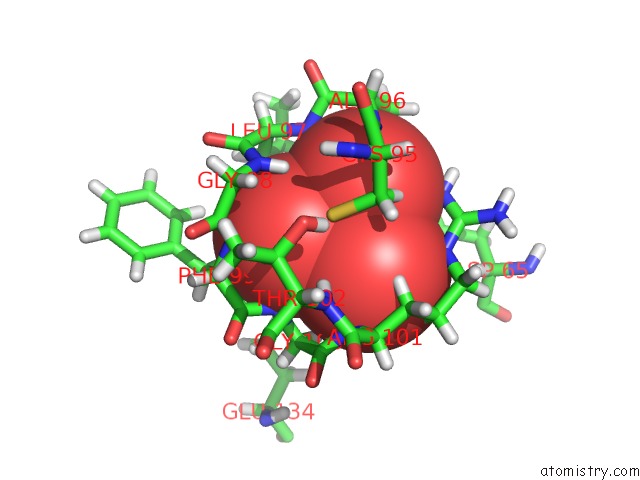
Mono view
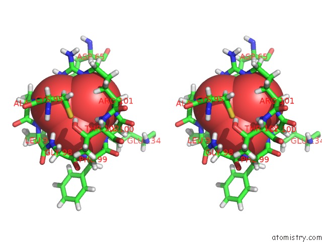
Stereo pair view
Mono view
Stereo pair view
A full contact list of Vanadium with other atoms in the V binding
site number 1 of Structure of Vhz Bound to Metavanadate within 5.0Å range:
|
Vanadium binding site 2 out of 2 in 4erc
Go back to
Vanadium binding site 2 out
of 2 in the Structure of Vhz Bound to Metavanadate
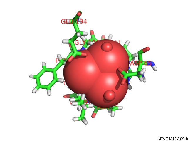
Mono view
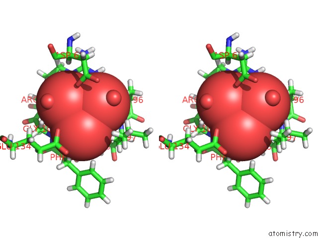
Stereo pair view
Mono view
Stereo pair view
A full contact list of Vanadium with other atoms in the V binding
site number 2 of Structure of Vhz Bound to Metavanadate within 5.0Å range:
|
Reference:
V.I.Kuznetsov,
A.C.Hengge,
S.J.Johnson.
New Aspects of the Phosphatase Vhz Revealed By A High-Resolution Structure with Vanadate and Substrate Screening. Biochemistry V. 51 9869 2012.
ISSN: ISSN 0006-2960
PubMed: 23145819
DOI: 10.1021/BI300908Y
Page generated: Fri Oct 11 19:39:44 2024
ISSN: ISSN 0006-2960
PubMed: 23145819
DOI: 10.1021/BI300908Y
Last articles
F in 7M00F in 7M01
F in 7M03
F in 7M02
F in 7LZU
F in 7LZW
F in 7LY8
F in 7LWG
F in 7LZV
F in 7LZF