Vanadium »
PDB 3omx-4zg4 »
4byf »
Vanadium in PDB 4byf: Crystal Structure of Human Myosin 1C in Complex with Calmodulin in the Pre-Power Stroke State
Enzymatic activity of Crystal Structure of Human Myosin 1C in Complex with Calmodulin in the Pre-Power Stroke State
All present enzymatic activity of Crystal Structure of Human Myosin 1C in Complex with Calmodulin in the Pre-Power Stroke State:
3.6.4.1;
3.6.4.1;
Protein crystallography data
The structure of Crystal Structure of Human Myosin 1C in Complex with Calmodulin in the Pre-Power Stroke State, PDB code: 4byf
was solved by
S.Munnich,
M.H.Taft,
S.Pathan-Chhatbar,
D.J.Manstein,
with X-Ray Crystallography technique. A brief refinement statistics is given in the table below:
| Resolution Low / High (Å) | 47.94 / 2.74 |
| Space group | P 1 21 1 |
| Cell size a, b, c (Å), α, β, γ (°) | 59.580, 158.450, 114.340, 90.00, 91.56, 90.00 |
| R / Rfree (%) | 18.3 / 23.67 |
Other elements in 4byf:
The structure of Crystal Structure of Human Myosin 1C in Complex with Calmodulin in the Pre-Power Stroke State also contains other interesting chemical elements:
| Magnesium | (Mg) | 3 atoms |
Vanadium Binding Sites:
The binding sites of Vanadium atom in the Crystal Structure of Human Myosin 1C in Complex with Calmodulin in the Pre-Power Stroke State
(pdb code 4byf). This binding sites where shown within
5.0 Angstroms radius around Vanadium atom.
In total 2 binding sites of Vanadium where determined in the Crystal Structure of Human Myosin 1C in Complex with Calmodulin in the Pre-Power Stroke State, PDB code: 4byf:
Jump to Vanadium binding site number: 1; 2;
In total 2 binding sites of Vanadium where determined in the Crystal Structure of Human Myosin 1C in Complex with Calmodulin in the Pre-Power Stroke State, PDB code: 4byf:
Jump to Vanadium binding site number: 1; 2;
Vanadium binding site 1 out of 2 in 4byf
Go back to
Vanadium binding site 1 out
of 2 in the Crystal Structure of Human Myosin 1C in Complex with Calmodulin in the Pre-Power Stroke State

Mono view
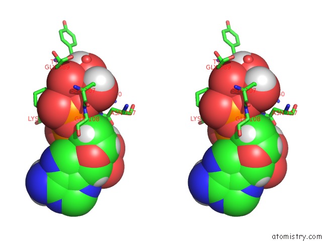
Stereo pair view
Mono view
Stereo pair view
A full contact list of Vanadium with other atoms in the V binding
site number 1 of Crystal Structure of Human Myosin 1C in Complex with Calmodulin in the Pre-Power Stroke State within 5.0Å range:
|
Vanadium binding site 2 out of 2 in 4byf
Go back to
Vanadium binding site 2 out
of 2 in the Crystal Structure of Human Myosin 1C in Complex with Calmodulin in the Pre-Power Stroke State
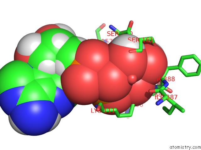
Mono view

Stereo pair view
Mono view
Stereo pair view
A full contact list of Vanadium with other atoms in the V binding
site number 2 of Crystal Structure of Human Myosin 1C in Complex with Calmodulin in the Pre-Power Stroke State within 5.0Å range:
|
Reference:
S.Munnich,
M.H.Taft,
D.J.Manstein.
Crystal Structure of Human Myosin 1C-the Motor in GLUT4 Exocytosis: Implications For Ca(2+) Regulation and 14-3-3 Binding. J.Mol.Biol. V. 426 2070 2014.
ISSN: ISSN 0022-2836
PubMed: 24636949
DOI: 10.1016/J.JMB.2014.03.004
Page generated: Tue Aug 19 08:07:13 2025
ISSN: ISSN 0022-2836
PubMed: 24636949
DOI: 10.1016/J.JMB.2014.03.004
Last articles
W in 1DV4W in 1FR3
W in 1GUG
W in 1H9R
W in 1H9K
W in 1H0H
W in 1FEZ
W in 1FKA
W in 1E3P
W in 1E18