Vanadium »
PDB 3omx-4zg4 »
3s3f »
Vanadium in PDB 3s3f: Crystal Structure of the Catalytic Domain of PTP10D From Drosophila Melanogaster with A Small Molecule Inhibitor Vanadate
Enzymatic activity of Crystal Structure of the Catalytic Domain of PTP10D From Drosophila Melanogaster with A Small Molecule Inhibitor Vanadate
All present enzymatic activity of Crystal Structure of the Catalytic Domain of PTP10D From Drosophila Melanogaster with A Small Molecule Inhibitor Vanadate:
3.1.3.48;
3.1.3.48;
Protein crystallography data
The structure of Crystal Structure of the Catalytic Domain of PTP10D From Drosophila Melanogaster with A Small Molecule Inhibitor Vanadate, PDB code: 3s3f
was solved by
L.L.Madan,
B.Gopal,
with X-Ray Crystallography technique. A brief refinement statistics is given in the table below:
| Resolution Low / High (Å) | 39.68 / 2.70 |
| Space group | P 31 2 1 |
| Cell size a, b, c (Å), α, β, γ (°) | 103.070, 103.070, 173.401, 90.00, 90.00, 120.00 |
| R / Rfree (%) | 23.8 / 27.2 |
Vanadium Binding Sites:
The binding sites of Vanadium atom in the Crystal Structure of the Catalytic Domain of PTP10D From Drosophila Melanogaster with A Small Molecule Inhibitor Vanadate
(pdb code 3s3f). This binding sites where shown within
5.0 Angstroms radius around Vanadium atom.
In total only one binding site of Vanadium was determined in the Crystal Structure of the Catalytic Domain of PTP10D From Drosophila Melanogaster with A Small Molecule Inhibitor Vanadate, PDB code: 3s3f:
In total only one binding site of Vanadium was determined in the Crystal Structure of the Catalytic Domain of PTP10D From Drosophila Melanogaster with A Small Molecule Inhibitor Vanadate, PDB code: 3s3f:
Vanadium binding site 1 out of 1 in 3s3f
Go back to
Vanadium binding site 1 out
of 1 in the Crystal Structure of the Catalytic Domain of PTP10D From Drosophila Melanogaster with A Small Molecule Inhibitor Vanadate
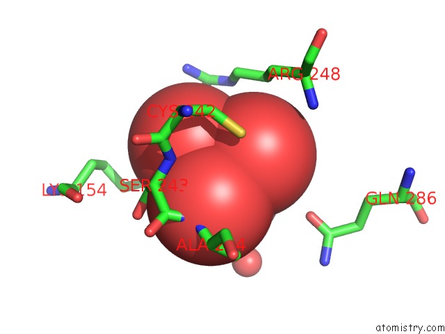
Mono view
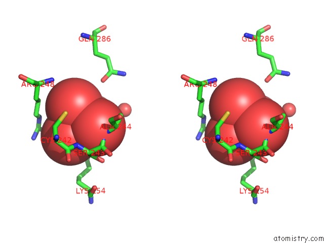
Stereo pair view
Mono view
Stereo pair view
A full contact list of Vanadium with other atoms in the V binding
site number 1 of Crystal Structure of the Catalytic Domain of PTP10D From Drosophila Melanogaster with A Small Molecule Inhibitor Vanadate within 5.0Å range:
|
Reference:
L.L.Madan,
B.Gopal.
Conformational Basis For Substrate Recruitment in Protein Tyrosine Phosphatase 10D Biochemistry V. 50 10114 2011.
ISSN: ISSN 0006-2960
PubMed: 22007620
DOI: 10.1021/BI201092Q
Page generated: Tue Aug 19 08:05:40 2025
ISSN: ISSN 0006-2960
PubMed: 22007620
DOI: 10.1021/BI201092Q
Last articles
W in 1DV4W in 1FR3
W in 1GUG
W in 1H9R
W in 1H9K
W in 1H0H
W in 1FEZ
W in 1FKA
W in 1E3P
W in 1E18