Vanadium »
PDB 1z12-3myh »
3gw8 »
Vanadium in PDB 3gw8: Crystal Structure of Phosphoglyceromutase From Burkholderia Pseudomallei with Vanadate and Glycerol
Enzymatic activity of Crystal Structure of Phosphoglyceromutase From Burkholderia Pseudomallei with Vanadate and Glycerol
All present enzymatic activity of Crystal Structure of Phosphoglyceromutase From Burkholderia Pseudomallei with Vanadate and Glycerol:
5.4.2.1;
5.4.2.1;
Protein crystallography data
The structure of Crystal Structure of Phosphoglyceromutase From Burkholderia Pseudomallei with Vanadate and Glycerol, PDB code: 3gw8
was solved by
Seattle Structural Genomics Center For Infectious Disease (Ssgcid),
with X-Ray Crystallography technique. A brief refinement statistics is given in the table below:
| Resolution Low / High (Å) | 50.00 / 1.93 |
| Space group | P 1 |
| Cell size a, b, c (Å), α, β, γ (°) | 45.175, 49.112, 62.590, 106.42, 91.18, 107.61 |
| R / Rfree (%) | 18.7 / 22.7 |
Vanadium Binding Sites:
The binding sites of Vanadium atom in the Crystal Structure of Phosphoglyceromutase From Burkholderia Pseudomallei with Vanadate and Glycerol
(pdb code 3gw8). This binding sites where shown within
5.0 Angstroms radius around Vanadium atom.
In total 4 binding sites of Vanadium where determined in the Crystal Structure of Phosphoglyceromutase From Burkholderia Pseudomallei with Vanadate and Glycerol, PDB code: 3gw8:
Jump to Vanadium binding site number: 1; 2; 3; 4;
In total 4 binding sites of Vanadium where determined in the Crystal Structure of Phosphoglyceromutase From Burkholderia Pseudomallei with Vanadate and Glycerol, PDB code: 3gw8:
Jump to Vanadium binding site number: 1; 2; 3; 4;
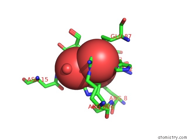
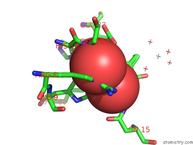
Vanadium binding site 1 out of 4 in 3gw8
Go back to
Vanadium binding site 1 out
of 4 in the Crystal Structure of Phosphoglyceromutase From Burkholderia Pseudomallei with Vanadate and Glycerol
Mono view
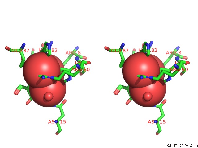
Stereo pair view
Mono view
Stereo pair view
A full contact list of Vanadium with other atoms in the V binding
site number 1 of Crystal Structure of Phosphoglyceromutase From Burkholderia Pseudomallei with Vanadate and Glycerol within 5.0Å range:
|
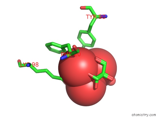
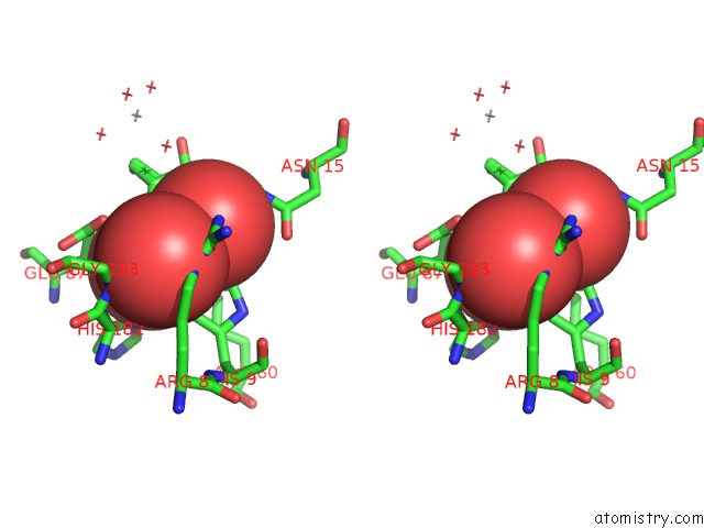
Vanadium binding site 2 out of 4 in 3gw8
Go back to
Vanadium binding site 2 out
of 4 in the Crystal Structure of Phosphoglyceromutase From Burkholderia Pseudomallei with Vanadate and Glycerol
Mono view
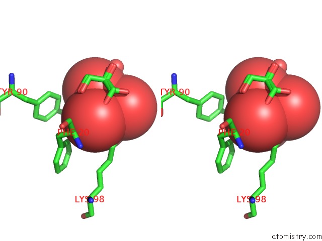
Stereo pair view
Mono view
Stereo pair view
A full contact list of Vanadium with other atoms in the V binding
site number 2 of Crystal Structure of Phosphoglyceromutase From Burkholderia Pseudomallei with Vanadate and Glycerol within 5.0Å range:
|
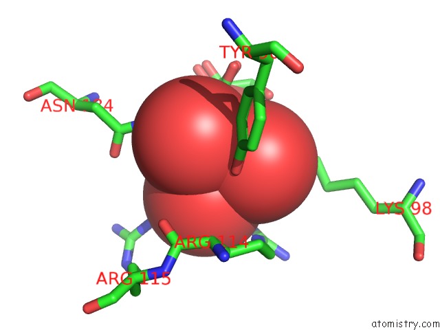
Vanadium binding site 3 out of 4 in 3gw8
Go back to
Vanadium binding site 3 out
of 4 in the Crystal Structure of Phosphoglyceromutase From Burkholderia Pseudomallei with Vanadate and Glycerol
Mono view
Stereo pair view
Mono view
Stereo pair view
A full contact list of Vanadium with other atoms in the V binding
site number 3 of Crystal Structure of Phosphoglyceromutase From Burkholderia Pseudomallei with Vanadate and Glycerol within 5.0Å range:
|
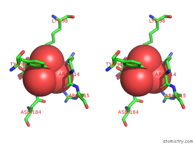
Vanadium binding site 4 out of 4 in 3gw8
Go back to
Vanadium binding site 4 out
of 4 in the Crystal Structure of Phosphoglyceromutase From Burkholderia Pseudomallei with Vanadate and Glycerol
Mono view
Stereo pair view
Mono view
Stereo pair view
A full contact list of Vanadium with other atoms in the V binding
site number 4 of Crystal Structure of Phosphoglyceromutase From Burkholderia Pseudomallei with Vanadate and Glycerol within 5.0Å range:
|
Reference:
D.R.Davies,
B.L.Staker,
J.A.Abendroth,
T.E.Edwards,
R.Hartley,
J.Leonard,
H.Kim,
A.L.Rychel,
S.N.Hewitt,
P.J.Myler,
L.J.Stewart.
An Ensemble of Structures of Burkholderia Pseudomallei 2,3-Bisphosphoglycerate-Dependent Phosphoglycerate Mutase. Acta Crystallogr.,Sect.F V. 67 1044 2011.
ISSN: ESSN 1744-3091
PubMed: 21904048
DOI: 10.1107/S1744309111030405
Page generated: Fri Oct 11 19:19:28 2024
ISSN: ESSN 1744-3091
PubMed: 21904048
DOI: 10.1107/S1744309111030405
Last articles
Zn in 9MJ5Zn in 9HNW
Zn in 9G0L
Zn in 9FNE
Zn in 9DZN
Zn in 9E0I
Zn in 9D32
Zn in 9DAK
Zn in 8ZXC
Zn in 8ZUF