Vanadium »
PDB 1z12-3myh »
3gp5 »
Vanadium in PDB 3gp5: Crystal Structure of Phosphoglyceromutase From Burkholderia Pseudomallei with 3-Phosphoglyceric Acid and Vanadate
Enzymatic activity of Crystal Structure of Phosphoglyceromutase From Burkholderia Pseudomallei with 3-Phosphoglyceric Acid and Vanadate
All present enzymatic activity of Crystal Structure of Phosphoglyceromutase From Burkholderia Pseudomallei with 3-Phosphoglyceric Acid and Vanadate:
5.4.2.1;
5.4.2.1;
Protein crystallography data
The structure of Crystal Structure of Phosphoglyceromutase From Burkholderia Pseudomallei with 3-Phosphoglyceric Acid and Vanadate, PDB code: 3gp5
was solved by
Seattle Structural Genomics Center For Infectious Disease (Ssgcid),
with X-Ray Crystallography technique. A brief refinement statistics is given in the table below:
| Resolution Low / High (Å) | 50.00 / 2.25 |
| Space group | P 1 |
| Cell size a, b, c (Å), α, β, γ (°) | 44.749, 48.983, 62.960, 105.46, 91.15, 107.42 |
| R / Rfree (%) | 17.1 / 23.4 |
Vanadium Binding Sites:
The binding sites of Vanadium atom in the Crystal Structure of Phosphoglyceromutase From Burkholderia Pseudomallei with 3-Phosphoglyceric Acid and Vanadate
(pdb code 3gp5). This binding sites where shown within
5.0 Angstroms radius around Vanadium atom.
In total 2 binding sites of Vanadium where determined in the Crystal Structure of Phosphoglyceromutase From Burkholderia Pseudomallei with 3-Phosphoglyceric Acid and Vanadate, PDB code: 3gp5:
Jump to Vanadium binding site number: 1; 2;
In total 2 binding sites of Vanadium where determined in the Crystal Structure of Phosphoglyceromutase From Burkholderia Pseudomallei with 3-Phosphoglyceric Acid and Vanadate, PDB code: 3gp5:
Jump to Vanadium binding site number: 1; 2;
Vanadium binding site 1 out of 2 in 3gp5
Go back to
Vanadium binding site 1 out
of 2 in the Crystal Structure of Phosphoglyceromutase From Burkholderia Pseudomallei with 3-Phosphoglyceric Acid and Vanadate
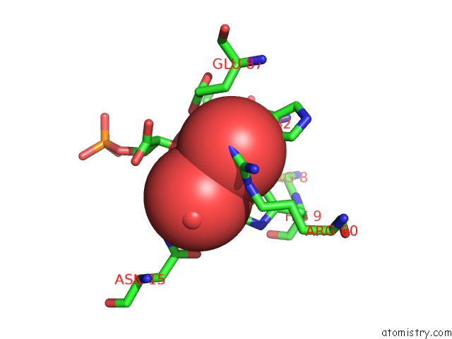
Mono view
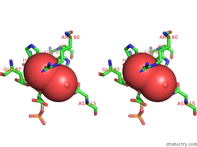
Stereo pair view
Mono view
Stereo pair view
A full contact list of Vanadium with other atoms in the V binding
site number 1 of Crystal Structure of Phosphoglyceromutase From Burkholderia Pseudomallei with 3-Phosphoglyceric Acid and Vanadate within 5.0Å range:
|
Vanadium binding site 2 out of 2 in 3gp5
Go back to
Vanadium binding site 2 out
of 2 in the Crystal Structure of Phosphoglyceromutase From Burkholderia Pseudomallei with 3-Phosphoglyceric Acid and Vanadate
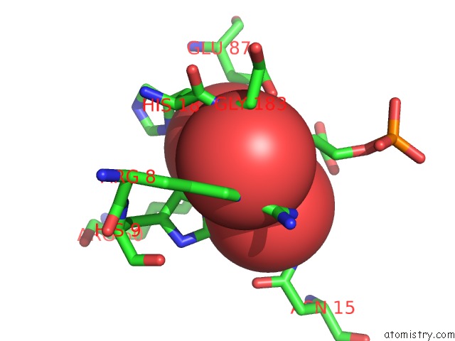
Mono view
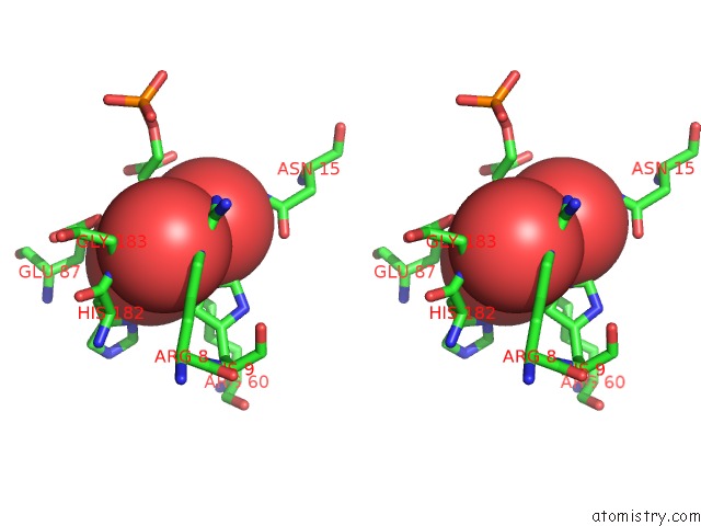
Stereo pair view
Mono view
Stereo pair view
A full contact list of Vanadium with other atoms in the V binding
site number 2 of Crystal Structure of Phosphoglyceromutase From Burkholderia Pseudomallei with 3-Phosphoglyceric Acid and Vanadate within 5.0Å range:
|
Reference:
D.R.Davies,
B.L.Staker,
J.A.Abendroth,
T.E.Edwards,
R.Hartley,
J.Leonard,
H.Kim,
A.L.Rychel,
S.N.Hewitt,
P.J.Myler,
L.J.Stewart.
An Ensemble of Structures of Burkholderia Pseudomallei 2,3-Bisphosphoglycerate-Dependent Phosphoglycerate Mutase. Acta Crystallogr.,Sect.F V. 67 1044 2011.
ISSN: ESSN 1744-3091
PubMed: 21904048
DOI: 10.1107/S1744309111030405
Page generated: Fri Oct 11 19:18:08 2024
ISSN: ESSN 1744-3091
PubMed: 21904048
DOI: 10.1107/S1744309111030405
Last articles
F in 4FS2F in 4FV0
F in 4FS1
F in 4FRJ
F in 4FRI
F in 4FOG
F in 4FQ4
F in 4FPH
F in 4FNZ
F in 4FOD