Vanadium »
PDB 1z12-3myh »
2v26 »
Vanadium in PDB 2v26: Myosin VI (Md) Pre-Powerstroke State (Mg.Adp.VO4)
Protein crystallography data
The structure of Myosin VI (Md) Pre-Powerstroke State (Mg.Adp.VO4), PDB code: 2v26
was solved by
J.Menetrey,
P.Llinas,
M.Mukherjea,
H.L.Sweeney,
A.Houdusse,
with X-Ray Crystallography technique. A brief refinement statistics is given in the table below:
| Resolution Low / High (Å) | 68.68 / 1.75 |
| Space group | P 21 21 21 |
| Cell size a, b, c (Å), α, β, γ (°) | 93.512, 93.845, 100.976, 90.00, 90.00, 90.00 |
| R / Rfree (%) | 20.2 / 22.7 |
Other elements in 2v26:
The structure of Myosin VI (Md) Pre-Powerstroke State (Mg.Adp.VO4) also contains other interesting chemical elements:
| Magnesium | (Mg) | 1 atom |
Vanadium Binding Sites:
The binding sites of Vanadium atom in the Myosin VI (Md) Pre-Powerstroke State (Mg.Adp.VO4)
(pdb code 2v26). This binding sites where shown within
5.0 Angstroms radius around Vanadium atom.
In total only one binding site of Vanadium was determined in the Myosin VI (Md) Pre-Powerstroke State (Mg.Adp.VO4), PDB code: 2v26:
In total only one binding site of Vanadium was determined in the Myosin VI (Md) Pre-Powerstroke State (Mg.Adp.VO4), PDB code: 2v26:
Vanadium binding site 1 out of 1 in 2v26
Go back to
Vanadium binding site 1 out
of 1 in the Myosin VI (Md) Pre-Powerstroke State (Mg.Adp.VO4)
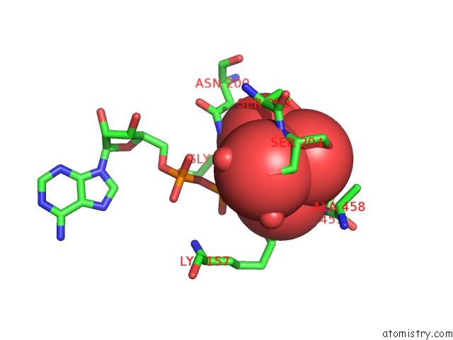
Mono view
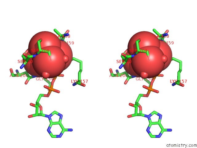
Stereo pair view
Mono view
Stereo pair view
A full contact list of Vanadium with other atoms in the V binding
site number 1 of Myosin VI (Md) Pre-Powerstroke State (Mg.Adp.VO4) within 5.0Å range:
|
Reference:
J.Menetrey,
P.Llinas,
M.Mukherjea,
H.L.Sweeney,
A.Houdusse.
The Structural Basis For the Large Powerstroke of Myosin VI. Cell(Cambridge,Mass.) V. 131 300 2007.
ISSN: ISSN 0092-8674
PubMed: 17956731
DOI: 10.1016/J.CELL.2007.08.027
Page generated: Fri Oct 11 19:15:19 2024
ISSN: ISSN 0092-8674
PubMed: 17956731
DOI: 10.1016/J.CELL.2007.08.027
Last articles
Cl in 5KORCl in 5KOL
Cl in 5KN1
Cl in 5KMU
Cl in 5KMD
Cl in 5KMB
Cl in 5KMA
Cl in 5KM9
Cl in 5KM8
Cl in 5KM5