Vanadium »
PDB 1z12-3myh »
2jhr »
Vanadium in PDB 2jhr: Crystal Structure of Myosin-2 Motor Domain in Complex with Adp- Metavanadate and Pentabromopseudilin
Protein crystallography data
The structure of Crystal Structure of Myosin-2 Motor Domain in Complex with Adp- Metavanadate and Pentabromopseudilin, PDB code: 2jhr
was solved by
R.Fedorov,
M.Boehl,
G.Tsiavaliaris,
F.K.Hartmann,
P.Baruch,
B.Brenner,
R.Martin,
H.J.Knoelker,
H.O.Gutzeit,
D.J.Manstein,
with X-Ray Crystallography technique. A brief refinement statistics is given in the table below:
| Resolution Low / High (Å) | 8.00 / 2.80 |
| Space group | C 2 2 21 |
| Cell size a, b, c (Å), α, β, γ (°) | 89.758, 150.464, 154.550, 90.00, 90.00, 90.00 |
| R / Rfree (%) | 21.7 / 26.5 |
Other elements in 2jhr:
The structure of Crystal Structure of Myosin-2 Motor Domain in Complex with Adp- Metavanadate and Pentabromopseudilin also contains other interesting chemical elements:
| Bromine | (Br) | 5 atoms |
| Magnesium | (Mg) | 1 atom |
Vanadium Binding Sites:
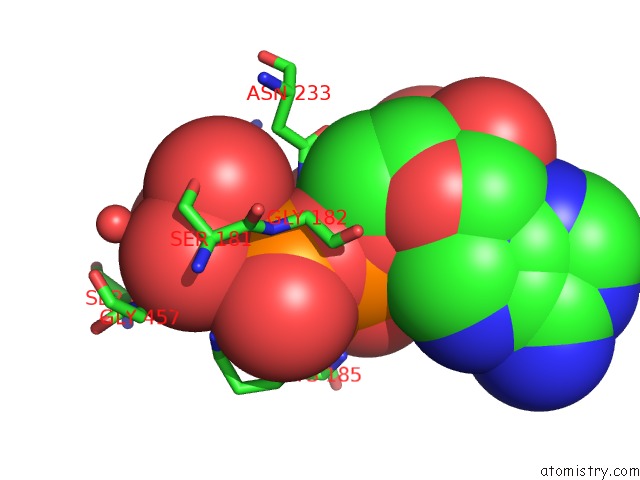
The binding sites of Vanadium atom in the Crystal Structure of Myosin-2 Motor Domain in Complex with Adp- Metavanadate and Pentabromopseudilin
(pdb code 2jhr). This binding sites where shown within
5.0 Angstroms radius around Vanadium atom.
In total only one binding site of Vanadium was determined in the Crystal Structure of Myosin-2 Motor Domain in Complex with Adp- Metavanadate and Pentabromopseudilin, PDB code: 2jhr:
In total only one binding site of Vanadium was determined in the Crystal Structure of Myosin-2 Motor Domain in Complex with Adp- Metavanadate and Pentabromopseudilin, PDB code: 2jhr:
Vanadium binding site 1 out of 1 in 2jhr
Go back to
Vanadium binding site 1 out
of 1 in the Crystal Structure of Myosin-2 Motor Domain in Complex with Adp- Metavanadate and Pentabromopseudilin
Mono view
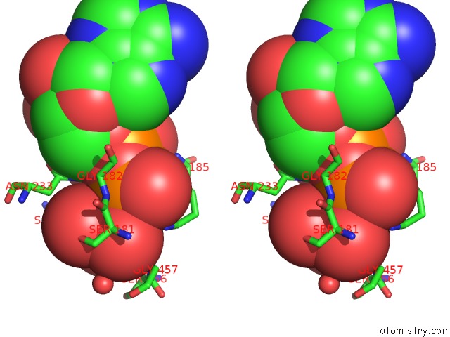
Stereo pair view
Mono view
Stereo pair view
A full contact list of Vanadium with other atoms in the V binding
site number 1 of Crystal Structure of Myosin-2 Motor Domain in Complex with Adp- Metavanadate and Pentabromopseudilin within 5.0Å range:
|
Reference:
R.Fedorov,
M.Bohl,
G.Tsiavaliaris,
F.K.Hartmann,
M.H.Taft,
P.Baruch,
B.Brenner,
R.Martin,
H.Knolker,
H.O.Gutzeit,
D.J.Manstein.
The Mechanism of Pentabromopseudilin Inhibition of Myosin Motor Activity. Nat.Struct.Mol.Biol. V. 16 80 2009.
ISSN: ISSN 1545-9993
PubMed: 19122661
DOI: 10.1038/NSMB.1542
Page generated: Fri Oct 11 19:12:08 2024
ISSN: ISSN 1545-9993
PubMed: 19122661
DOI: 10.1038/NSMB.1542
Last articles
Cl in 7ZYDCl in 7ZXV
Cl in 7ZWY
Cl in 7ZX4
Cl in 7ZXD
Cl in 7ZX2
Cl in 7ZWX
Cl in 7ZWT
Cl in 7ZWU
Cl in 7ZW2