Vanadium »
PDB 1z12-3myh »
2gso »
Vanadium in PDB 2gso: Structure of Xac Nucleotide Pyrophosphatase/Phosphodiesterase in Complex with Vanadate
Enzymatic activity of Structure of Xac Nucleotide Pyrophosphatase/Phosphodiesterase in Complex with Vanadate
All present enzymatic activity of Structure of Xac Nucleotide Pyrophosphatase/Phosphodiesterase in Complex with Vanadate:
3.6.1.9;
3.6.1.9;
Protein crystallography data
The structure of Structure of Xac Nucleotide Pyrophosphatase/Phosphodiesterase in Complex with Vanadate, PDB code: 2gso
was solved by
J.G.Zalatan,
T.D.Fenn,
A.T.Brunger,
D.Herschlag,
with X-Ray Crystallography technique. A brief refinement statistics is given in the table below:
| Resolution Low / High (Å) | 50.00 / 1.30 |
| Space group | P 21 21 21 |
| Cell size a, b, c (Å), α, β, γ (°) | 66.040, 78.776, 129.686, 90.00, 90.00, 90.00 |
| R / Rfree (%) | 17.2 / 19.3 |
Other elements in 2gso:
The structure of Structure of Xac Nucleotide Pyrophosphatase/Phosphodiesterase in Complex with Vanadate also contains other interesting chemical elements:
| Zinc | (Zn) | 4 atoms |
Vanadium Binding Sites:
The binding sites of Vanadium atom in the Structure of Xac Nucleotide Pyrophosphatase/Phosphodiesterase in Complex with Vanadate
(pdb code 2gso). This binding sites where shown within
5.0 Angstroms radius around Vanadium atom.
In total 2 binding sites of Vanadium where determined in the Structure of Xac Nucleotide Pyrophosphatase/Phosphodiesterase in Complex with Vanadate, PDB code: 2gso:
Jump to Vanadium binding site number: 1; 2;
In total 2 binding sites of Vanadium where determined in the Structure of Xac Nucleotide Pyrophosphatase/Phosphodiesterase in Complex with Vanadate, PDB code: 2gso:
Jump to Vanadium binding site number: 1; 2;
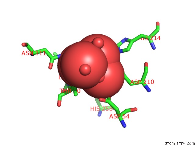
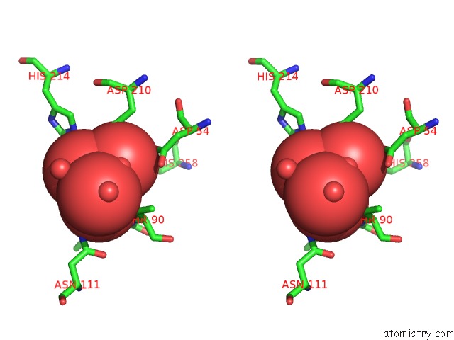
Vanadium binding site 1 out of 2 in 2gso
Go back to
Vanadium binding site 1 out
of 2 in the Structure of Xac Nucleotide Pyrophosphatase/Phosphodiesterase in Complex with Vanadate

Mono view

Stereo pair view
Mono view
Stereo pair view
A full contact list of Vanadium with other atoms in the V binding
site number 1 of Structure of Xac Nucleotide Pyrophosphatase/Phosphodiesterase in Complex with Vanadate within 5.0Å range:
|
Vanadium binding site 2 out of 2 in 2gso
Go back to
Vanadium binding site 2 out
of 2 in the Structure of Xac Nucleotide Pyrophosphatase/Phosphodiesterase in Complex with Vanadate
Mono view
Stereo pair view
Mono view
Stereo pair view
A full contact list of Vanadium with other atoms in the V binding
site number 2 of Structure of Xac Nucleotide Pyrophosphatase/Phosphodiesterase in Complex with Vanadate within 5.0Å range:
|
Reference:
J.G.Zalatan,
T.D.Fenn,
A.T.Brunger,
D.Herschlag.
Structural and Functional Comparisons of Nucleotide Pyrophosphatase/Phosphodiesterase and Alkaline Phosphatase: Implications For Mechanism and Evolution Biochemistry V. 45 9788 2006.
ISSN: ISSN 0006-2960
PubMed: 16893180
DOI: 10.1021/BI060847T
Page generated: Fri Oct 11 19:09:43 2024
ISSN: ISSN 0006-2960
PubMed: 16893180
DOI: 10.1021/BI060847T
Last articles
Zn in 9MJ5Zn in 9HNW
Zn in 9G0L
Zn in 9FNE
Zn in 9DZN
Zn in 9E0I
Zn in 9D32
Zn in 9DAK
Zn in 8ZXC
Zn in 8ZUF