Vanadium »
PDB 1z12-3myh »
1z12 »
Vanadium in PDB 1z12: Crystal Structure of Bovine Low Molecular Weight Ptpase Complexed with Vanadate
Enzymatic activity of Crystal Structure of Bovine Low Molecular Weight Ptpase Complexed with Vanadate
All present enzymatic activity of Crystal Structure of Bovine Low Molecular Weight Ptpase Complexed with Vanadate:
3.1.3.2; 3.1.3.48;
3.1.3.2; 3.1.3.48;
Protein crystallography data
The structure of Crystal Structure of Bovine Low Molecular Weight Ptpase Complexed with Vanadate, PDB code: 1z12
was solved by
M.Zhang,
M.Zhou,
R.L.Van Etten,
C.V.Stauffacher,
with X-Ray Crystallography technique. A brief refinement statistics is given in the table below:
| Resolution Low / High (Å) | 20.00 / 2.20 |
| Space group | C 1 2 1 |
| Cell size a, b, c (Å), α, β, γ (°) | 95.300, 43.400, 41.200, 90.00, 113.40, 90.00 |
| R / Rfree (%) | n/a / n/a |
Vanadium Binding Sites:
The binding sites of Vanadium atom in the Crystal Structure of Bovine Low Molecular Weight Ptpase Complexed with Vanadate
(pdb code 1z12). This binding sites where shown within
5.0 Angstroms radius around Vanadium atom.
In total only one binding site of Vanadium was determined in the Crystal Structure of Bovine Low Molecular Weight Ptpase Complexed with Vanadate, PDB code: 1z12:
In total only one binding site of Vanadium was determined in the Crystal Structure of Bovine Low Molecular Weight Ptpase Complexed with Vanadate, PDB code: 1z12:
Vanadium binding site 1 out of 1 in 1z12
Go back to
Vanadium binding site 1 out
of 1 in the Crystal Structure of Bovine Low Molecular Weight Ptpase Complexed with Vanadate
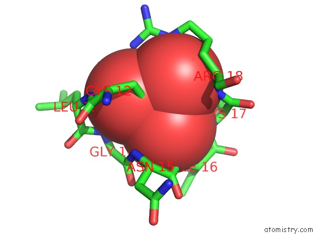
Mono view
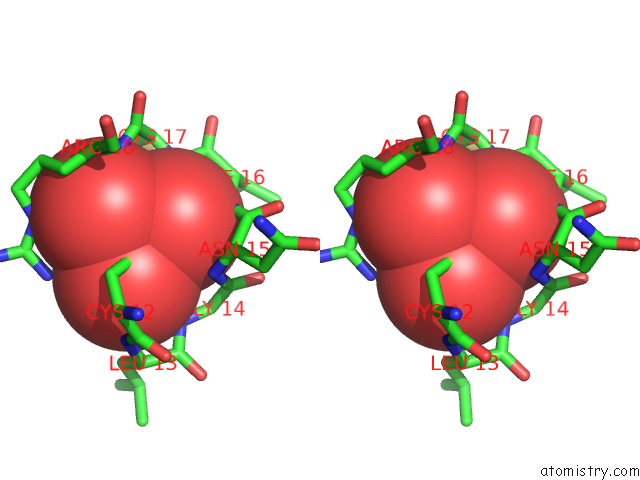
Stereo pair view
Mono view
Stereo pair view
A full contact list of Vanadium with other atoms in the V binding
site number 1 of Crystal Structure of Bovine Low Molecular Weight Ptpase Complexed with Vanadate within 5.0Å range:
|
Reference:
M.Zhang,
M.Zhou,
R.L.Van Etten,
C.V.Stauffacher.
Crystal Structure of Bovine Low Molecular Weight Phosphotyrosyl Phosphatase Complexed with the Transition State Analog Vanadate Biochemistry V. 36 15 1997.
ISSN: ISSN 0006-2960
PubMed: 8993313
DOI: 10.1021/BI961804N
Page generated: Fri Oct 11 19:08:26 2024
ISSN: ISSN 0006-2960
PubMed: 8993313
DOI: 10.1021/BI961804N
Last articles
Au in 5CVLAu in 5E5F
Au in 4B8B
Au in 5DNN
Au in 5CCW
Au in 4ZFP
Au in 4OVB
Au in 4ZGH
Au in 4Y2I
Au in 4Y2M